We all enjoy and appreciate colours, beauty and brightness of the world around us. Would it be possible for any living being to survive even a day without having a vision or sight? The sight is the most important sense that we humans have and it not only helps us in collecting visual information from the outside world, but also helps us in expressing our emotions and feelings. All the living beings in this world collect visual information from the outside world with the help of the Eyes. Be it watching a movie, riding a vehicle, enjoying a scenic beauty or having a face-to-face communication with friends or with some we love. The excellence of sense of site that we humans have has tempted every individual to reply on it for the pursuit of food and safety. It would be really difficult for us to find our way around, or to steer our self on the road for even a short distance without the help of our Eyes. Apart from the visual information, our knowledge about the world comes through our Eyes.
The Eyes are a pair of organs that are situated in the head through which we humans and vertebrate animals see. An Eye is basically an extraordinary complex organ that allows us to see.
All the parts of our Eyes are very delicate, so our bodies protect them in many ways. Our Eyeballs are situated inside the orbit (Eye socket) in the skull, where further they are surrounded by bones. The orbit comprises of seven bones that helps us to protect our Eyes from injury. The seven bones include Sphenoid, Frontal, Ethmoid, Lachrimal, Maxillary, Palatine and Zygomatic. These seven bones get converged and form a pyramid-shaped structure. Within these Eye sockets or the orbit our Eyeballs rest. There is a layer of fat within the socket which helps the Eyes to move smoothly within the orbit.
Our Eyeball consists of three layers:
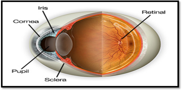
The outer layer is formed by the sclera (the white part) and the cornea (the clear dome over the iris).
The middle layer holds the primary blood supply for the Eye and it contains the iris (the pigmented part) and the pupil (the black circular opening in the iris that allows the light in).
The inner layer comprises the retina.

The outer layer of our Eyeball is white and tough, with an opaque membrane known as the sclera. The sclera is opaque, and many blood vessels and nerves run through it, including the optical nerve. The cornea is the transparent front window of the Eye, which is thick, circular structure that covers the lens. The cornea is clear, thin and a dome-shaped tissue. It basically covers the pupil and the iris. The cornea also covers one-sixth of the Eye. The cornea and the sclera meet together at a point known as the limbus, which contains blood vessels.
The most noticeable part of our Eyes is the iris and the pupil. The coloured membrane of our Eyes is known as the iris. The iris is situated between the cornea and the lens. It is primarily a coloured ring of tissues that lie beneath the cornea and can be in range of colours from pale blue to brown, which depends on the genetics. The circular black hole situated in the centre of iris is known as the pupil. The pupil acts like a camera aperture and allows the light to enter the Eye. The pupil works in the same way that as of a camera, as it adjusts according to the lighting conditions. In very bright conditions, the pupil closes down and reduces the amount of light to enter the Eye, thus protecting the delicate nerves from being damaged. In dark conditions, reverse action takes place. The size of the pupil changes automatically according to the lighting conditions, in order to control the amount of light entering the Eye.
The inner layer, which lines back two-thirds of the Eyeball is known as the retina. The retina is made of millions of visual cells, which are connected by the optic nerve to the brain. The sight that we see is the result of electrical impulses that are sent to the brain by the retina. The retina consists of two layers: the retinal pigment epithelium – it lies between the sensory retina and the wall of the Eye; the sensory retina – it contains the nerve cells that help in processing visual information that are sent to the brain.
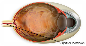
Along with numerous blood vessels, our Eyes also consist of the optic nerve, which runs from the back of our Eyeball, through an opening in the orbit known as the optic foramen. It also connects to the brain and acts as a conduit, which transmits visual information to the brain. The other nerves in the Eye carry non-visual information and send messages related to pain, stress or help to control the basic motor activity within the Eye.

Though the Eyelids are often ignored, they do play a major role in protecting our Eyes. The Eyelids protect the surface of our Eyes from dust, foreign objects and scratches. The Eyelids also lubricate the surface of our Eyes. The Eyelids carry secretions from various glands across the Eye when we blink.
The Eyelids have several layers:
-
To provide stability there is a fibrous layer.
-
For controlling the opening and closing of the Eyes there is also a layer of muscle that controls it. If the surface of the Eye is under an attack, this layer acts quickly by shutting the Eyelid and by protecting the surface of the Eyes by using a reflex mechanism.
-
The Eyelids also contain a layer of skin which contains glands that basically acts as a filter to stop large foreign objects from entering the Eye.
-
The conjunctiva is the most important layer as it is mucous membrane that connects our Eyeball to the Eyelid and the Eyeball to the orbit.
In and around our Eyelids several glands provide the tears. The tears consist of around 99 percent water, and they protect the delicate cells on the cornea, along with keeping the cornea moist. When we blink, the tears drain away through the puncta lacrimal, which is a small opening situated in the inner corner of our Eyelids.
The Eyeball consists of three chambers of fluid:
-
Anterior chamber: The front part of the Eye, between the iris and the cornea is the anterior chamber.
-
Posterior chamber: Situated between the lens and the iris is the posterior chamber. The lens situated behind the iris is clear, and the light passes through the lens. The lens is situated in a place by small tissue fibers, which extend from the inner wall of the Eye. There are also small muscles, which are attached to lens that allow it to change it shape to focus on the objects that are at a varying distance.
-
Vitreous chamber: the vitreous chamber is situated between the back of the Eye and the lens. The inner wall of the back two thirds of the chamber is filled with special layers of cells, which convert the light into nerve impulses. The macula which is situated near the centre of the retina provides a detailed and a sharp vision for focussing on what is in front of us. The rest of the retina provides a peripheral vision which allows us to see shapes but not the details.
-
Myopia (short-sightedness): It is a defect more common in young people who see with the help of spectacles. In this disorder, it is difficult for the person to see distant objects clearly, although reading a book is possible for them if it is kept close. Myopia is basically the result of a very strong refraction and the person dealing with it should go for an Eye check at least once in a year.
-
Hypermetropia (Long-sightedness): in this defect the person is able to see distance objects clearly, but faces difficulty to see the details of the nearby objects. The person dealing with this problem finds reading a strain, and straining while reading may also lead to headache. In this condition, the power of the lens is not sufficient to focus the near objects clearly on the retina.
The other disorders associated with the malfunctioning of the Eyes are:
-
Astigmatism: It is a defect that causes problem to properly focus the light on the retina. It generally causes blurry vision.
-
Black Eye: Due to an injury to the face the area around the Eye swells and gets discoloured.
-
Cataract: It is clouding of the lens, which restricts the passage of the light through the lens.
-
Hyphema: It is caused mostly due to trauma, leads to bleeding into the front of the Eye, behind the cornea.
-
Keratitis: it is the inflammation or the infection of the cornea, which occurs after the germs enter a corneal abrasion.
-
Diplopia: Seeing double is known as diplopia and is usually caused due to serious conditions.
G Kowledge of | 0 Comments >>
0 Comments
Leave Comment
Your email address will not be published. Required fields are marked.